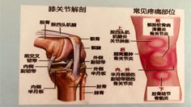
Unique features of thoracic vertebrae
Ribs, thinner discs, narrower canals
Identification of thoracic vertebrae, typical thoracic vertebrae, atypical thoracic vertebrae
First thoracic vertebra, ninth thoracic vertebra, tenth thoracic vertebra, eleventh thoracic vertebra, twelfth thoracic vertebra, thoracic joint, thoracic ligament
Anterior longitudinal ligament, posterior longitudinal ligament, intertransverse process ligament, cystic ligament, ligamentum flavum, interspinous ligament, supraspinatus ligament
Ligaments unique to the thoracic spine, thoracic muscles, external back muscles, and internal back muscles
Blood supply to thoracic vertebrae
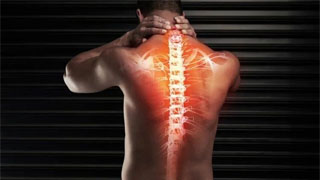
The thoracic vertebrae, also known as the dorsal vertebrae, consist of 12 vertebrae located behind the cervical vertebrae. These vertebrae are named T1 to T12 (or D1 to D12) from top to bottom.
If you palpate from top to bottom on the midline of the neck, you will notice a sudden protrusion or lump in the lower part of the neck. This protrusion is the posterior spinous process of the C7 vertebra, which protrudes due to its larger size. C7 is connected to T1.
Similarly, the lowest vertebra T12 in the thoracic spine is connected to the first vertebra of L1 or lumbar spine. There are ten thoracic vertebrae connecting the two.
The thoracic vertebrae help support the torso and chest area of the body, and provide attachment points for every rib except for the two ribs at the bottom of the chest.
Like most other vertebrae, the thoracic vertebrae are circular. The bone arch protrudes from the posterior part of each vertebral body, forming a hollow protective space containing the spinal cord. The thoracic facet joints are paired at the posterior portion of each vertebra, allowing for limited spinal movement.
Unique features of thoracic vertebrae
Compared with cervical vertebrae, thoracic vertebrae have the following unique characteristics
Ribs
Each thoracic vertebra is accompanied by a pair of ribs. The ribs are connected to the spine backwards and to the sternum forwards.
The first, tenth, and twelfth pairs of ribs are only connected to the vertebrae they are named after.
The second to tenth ribs are hinged to their named vertebral bodies, intervertebral discs, and the vertebrae above them.
Each pair of ribs is also connected to the anterior surface joint of its designated vertebral transverse process.
The thoracic vertebrae and thorax provide stability and protect important organs of the chest.
Thinner intervertebral discs
The intervertebral disc is a cushion between two vertebrae that serves as a shock absorber. The intervertebral discs of the thoracic spine are thinner than those of the cervical or lower spine.
Narrower canals
The spinal canal is the narrowest among the thoracic vertebrae. Compared to lumbar vertebrae with wider tubes, chest injuries are one of the reasons for more spinal cord injuries.
Identification of thoracic vertebrae
The thoracic spine is identified by the presence of rib surfaces on the lateral surface of the vertebral body. The rib surface of each vertebra is connected to the corresponding rib. There may be two or only one rib on each side. There are twelve thoracic vertebrae [typical and atypical thoracic vertebrae], among which the 2nd to 8th vertebrae are typical, and the remaining 5 vertebrae, namely the 1st, 9th, 10th, 11th, and 12th vertebrae, are atypical.
Typical thoracic vertebrae
A typical thoracic vertebral body is heart-shaped, with approximately the same size on the left, right, and anterior posterior sides. There are two ribs (half sides) on each side. The upper rib joint surface is relatively large, located at the upper edge of the body, close to the pedicle. They are hinged to the heads of the ribs corresponding to the numbers. The lower rib joint surface is relatively small and located at the lower edge in front of the lower vertebral incision. They are connected to the next rib.
The vertebral foramen is relatively small and circular. The pedicle of the vertebral arch extends directly backwards. The supravertebral incisions are shallow, while the infravertebral incisions are deep and distinct. The thin sheets overlap each other from above.
The superior articular process protrudes upwards from the junction of the pedicle and vertebral plate. The joint surface is flat and oriented backwards, slightly laterally and upwards. This direction allows for rotational movement of the spine.
Fusion of inferior articular process and vertebral plate. Their joints face forward and slightly downward and inward.
The transverse process is large and extends laterally and posteriorly from the junction of the pedicle and vertebral plate. Each protrusion has a small surface near its tip, which is used to connect with the nodules of the corresponding rib. In the upper 6 vertebrae, the rib facets on the transverse process are concave, facing forward and laterally. In other cases, the facets are flat and face upwards, laterally, and slightly forward.
The spinous process is very long and extends downwards and backwards. The 5th to 9th spinous processes are the longest, more vertical, and overlap with each other. The inclination direction of the spinous process of the vertebrae near the junction of the cervical and lumbar vertebrae is relatively small.
enclosure
The upper and lower boundaries of the body - the anterior and posterior longitudinal ligaments.
The upper and lower parts of the anterior surface - the ligamentum flavum or ligamentum flavum.
Transverse process
Lateral transverse rib ligament (tip), superior transverse rib ligament (lower edge), inferior transverse rib ligament (anterior surface), transverse muscle (upper and lower boundaries), levator rib muscle (posterior surface).
Spinous process
Supraspinatus ligament and interspinous ligament
Several muscles, including trapezius, rhombus, latissimus dorsi, posterior serrations, and inferior serrations
Atypical thoracic vertebrae
First thoracic vertebra
T1 vertebrae are similar to cervical vertebrae. It's wide, not heart-shaped. Its upper surface has a lip shape laterally and tilts forward. The rib surface on the body has been completed. It is connected to the head of the first rib. The articular surface of the lower rib is the "half face" of the second rib.
The spine is thick, long, and almost horizontal. Like the cervical spine, the upper vertebrae have distinct incisions.
Ninth thoracic vertebra
It is similar to a typical thoracic spine, except that the body only has the upper rib surface (half face). The lower rib surface does not exist.
Tenth thoracic vertebra
It is similar to a typical thoracic spine, except that each side of the vertebral body has a complete upper rib facet that extends to the root of the pedicle.
Eleventh thoracic vertebra
Each side of the body has a large rib facet that extends to the upper part of the pedicle.
The transverse process is very small and there is no articular surface. Sometimes it is difficult to distinguish between T10 vertebrae and T11 vertebrae.
Twelfth thoracic vertebra
The shape of the body, pedicle, transverse process, and spine is similar to that of the lumbar spine. However, there is a rib facet on each side of the body, which is located more in the lower part of the pedicle than on the body.
The transverse process is small and lacks small facets, but there are upper nodules, lower nodules, and lateral nodules.
The lower articular surface is of the lumbar spine type and points laterally (outward rotation), but the upper articular surface is of the thoracic spine type.
Thoracic joint
There are also associations and small joints in other areas of the spine. The rib joint is unique to the thoracic spine.
Symphony
Adjacent vertebral bodies are connected by intervertebral discs made of fibrocartilage.
apophyseal joint
These are synovial joints formed by connecting the superior and inferior articular processes of adjacent vertebrae.
The following joints are unique to the thoracic spine.
Rib vertebral joint
Each rib joint is composed of a rib head, which is connected by:
The upper rib joint of the corresponding vertebra, the lower rib joint of the upper vertebra, and the intervertebral disc separate the two vertebrae
The joint is stabilized by the intra-articular ligament of the rib head, which connects the rib head to the intervertebral disc. These joints can only undergo slight sliding movements.
Transverse rib joint
The transverse rib joint is formed by the joint between the thoracic transverse process and adjacent rib nodules. They exist in all vertebrae except T11 and T12.
Thoracic ligament
The presence of a large number of ligaments enhances the strength of the thoracic spine.
Anterior longitudinal ligament
The anterior longitudinal ligament extends along the anterior surface of the vertebral body from the occipital bone to the sacrum. The anterior longitudinal ligament in the chest area is thick and narrow. Ligaments are firmly attached to the edge of the vertebral body.
It is thicker and slightly narrower on the vertebral body, and thinner but slightly wider on the intervertebral disc. It is also loosely attached to the intervertebral disc ring.
It is a three-layer ligament - superficial, intermediate, and deep.
Posterior longitudinal ligament
The posterior longitudinal ligament originates from the posterior part of the occipital bone, is continuous with the periosteum, passes through the posterior surface of the vertebral body, and descends to the coccyx. It is located within the spinal canal.
Intertransverse process ligament
These ligaments are located between the transverse processes. In the chest area, they are closely connected to the deep muscles of the back.
Cystic ligament
The capsule ligament attaches to the joint edge of the articular process. The direction of the fibers is perpendicular to the small joints, and the fibers in the chest and waist regions are stronger than those in the neck region.
Yellow ligament
The ligamentum flavum, also known as the yellow ligament, connects the anterior lower edge of the vertebral plate to the posterior upper edge of the lower vertebral plate. The yellow ligament in the chest area is thicker. This ligament is mainly composed of elastic fibers, whose elasticity helps maintain an upright posture and assists in the recovery of the spine after flexion.
Interspinous ligament
The interspinous ligament connects adjacent spinous processes and slopes from the anterior inferior aspect of the superior spinous process to the posterior superior aspect of the spinous process
Supraspinatus ligament
This sturdy ligament is a strong fibrous cord that connects the spinous process tips from C7 to the sacrum together.
In the cervical spine, the interspinous and supraspinal ligaments thicken and combine to form the cervical ligament
Ligaments unique to thoracic vertebrae
Radiating ligament of rib head
The ligament extends fan-shaped outward from the head of the rib to the body of the two vertebrae and intervertebral disc.
Transverse rib ligament
It connects the rib neck and transverse process.
Lateral transverse rib ligament
It extends from the transverse process to the rib nodules.
Superior transverse rib ligament
It runs from the upper edge of the rib neck to the transverse process of the vertebrae above it.
Thoracic muscles
There are two types of muscles in the back
External factors responsible for limb and respiratory movements
The intrinsic factors for maintaining posture and performing spinal movements.
External muscles of the back
The external muscles of the back include superficial muscles and intermediate muscles.
shallow
The trapezius, latissimus dorsi, levator scapula, and rhombus muscles connect upper limb and trunk movements to move limbs
middle
Upper and lower serratus muscles, thin and shallow respiratory muscles
Internal muscles in the back
Internal or deep back muscles maintain posture and control the movement of the spine.
The deep back muscles can be divided into shallow, middle, and deep layers
surface
Head muscles, neck muscles.
Middle layer [erector spinae muscle]
Iliopsoas muscle, longest muscle, spinal muscle. The main extensor muscles of the spine.
Deep [transverse spinal muscle]
Hemispinae muscle, multifidus muscle, rotator
In addition, there are also the following muscles.
Intervertebral bodies connecting spinous processes
Connecting the transverse processes between them
The anterior muscle tissue is located in the cervical and lumbar regions, not the chest region.
Blood supply to thoracic vertebrae
The branch of the posterior intercostal artery is the main blood supplier to the thoracic spine.
The first two intercostal arteries are branches of the subclavian artery. The remaining intercostal arteries are branches of the thoracic aorta.
The intercostal artery extends to the periosteal artery and equatorial artery. These further branches into anterior and posterior root canal branches.
There is a venous plexus inside the spinal canal and another venous plexus outside the spinal canal. There are no valves on either side of the nerve plexus. This allows blood to move according to pressure gradients. The blood ultimately flows into the segmental veins of the trunk.