The study of anatomy is one of the difficulties that many rehabilitation practitioners cannot avoid. With numerous muscle movements, anatomical locations, complex functional decomposition, and the relationship between active muscles, coordinating muscles, and antagonistic muscles, it is not enough to complete muscle learning. There are more complex nervous system innervation behind muscles. It's like an endless sea of learning and an endless sea of hardship.
What's even more embarrassing is that many friends stay up all night memorizing their nerves for a month, only to find out in the end that they don't need it!
However, what we want to teach you now is to take the radial nerve as an example and "the the Belt and Road Initiative" to see how to assess and rehabilitate the problem of radial nerve entrapment from a unique, interesting and detailed perspective, from the perspective of practicality and ease of use.
The radial nerve is located close to the shaft at the middle and lower one-third of the humerus, where it is susceptible to injury during humeral fractures. Excessive growth of callus and anterior dislocation of the radial head can compress the radial nerve. Carelessness during surgery can also damage this nerve. The radial nerve is composed of fibers from the neck 5-8 and chest 1 nerve roots, and is a continuation of the posterior bundle of the brachial plexus.
In the upper arm, the radial nerve exits the armpit and follows the deep brachial artery from the inner side of the upper arm, passing between the long and inner heads of the triceps brachii to the back of the upper arm. It then descends through the radial nerve groove of the humerus between the inner and outer heads of the triceps brachii, and when it reaches the outer side of the elbow, it divides into shallow and deep branches before entering the forearm. The radial nerve branches successively in the upper arm, innervating the triceps brachii, brachioradialis muscle, and extensor carpi radialis.
Why do we still need to learn neuroscience
First, let's ask a question - why do we still need to learn neurology when it's so difficult?
Answer: In one sentence, all structures are innervated by nerves!
This may seem like nonsense, and many students have heard their teachers repeat it countless times during their college years. However, this sentence should be interpreted as follows:
Since all structures are innervated by nerves, all problems that occur in our bodies, clients, or patients may also be accompanied by neurological issues.
Since all structures are innervated by nerves, we cannot ignore the role of nerves in any treatment or therapy - if you prick a needle, it may reach a nerve fiber, if you use a technique, it may reach a nerve outlet, if you do stretching or rehabilitation, it may and will definitely affect the nerves and work through them.
Sometimes, we find that some problems, we may not have done anything, and the other party miraculously heals themselves - at this time, perhaps the nerves are "secretly helping you";
Similarly, sometimes we find that we have done a lot of processing, but many problems have not been solved, which may also be due to the nerves playing tricks in the dark.
The radial nerve is a branch of the brachial plexus nerve, and it is said to be the thickest, longest, and largest among the branches of the brachial plexus.
The brachial plexus is mainly composed of the anterior branches of the 5th to 8th cervical nerves and the anterior branch of the first thoracic nerve. It passes through the space between the scalene muscles, travels behind and above the subclavian artery, and enters the armpit through the posterior part of the clavicle.
Because there are too many branches of the brachial plexus, we teach everyone a number memorization method: "Five roots/Three stems/Six strands/Three bundles/Fourteen branches".
Five roots - Five nerve roots (C5 \ C6 \ C7 \ C8 \ T1);
Three stems - five stems combined to form the upper, middle, and lower three stems;
Six shares - Three stems issue two shares before and after, 3 * 2=6 shares;
Three bundles - six strands combined into three bundles, namely the lateral bundle, posterior bundle, and medial bundle;
Fourteen Branches - There are a total of fourteen main branches of the brachial plexus;
Since the radial nerve is part of the brachial plexus, which passes out from the side of the cervical spine, the first point of compression is
1. Cervical spine.
Misalignment of the cervical spine can easily cause compression of the cervical or brachial plexus nerves. Therefore, if there is a straightening of the cervical curvature, cervical osteophyte formation, or other conditions, the reduction and rehabilitation training of the cervical spine must be considered during the rehabilitation process. (This part is quite complex, especially the cervical reduction technique involves many contraindications, which we will not elaborate on here)
2. Diagonal muscle.
As we mentioned earlier, the brachial plexus nerve penetrates through the gap between the anterior and middle scalene muscles. Therefore, if the scalene muscle becomes tense due to prolonged posture such as lowering the head, it will compress the brachial plexus nerve here.
The release technique of the scalene muscle should be performed on the lateral side of the cervical spine.
Then, the radial nerve continues to descend and wraps around the armpit below the clavicle. This can easily cause compression here because there is one - the pectoralis minor muscle.
3. Pectoral muscles.
Similarly, due to long-term desk work, the shoulders are constantly in a forward rotation state. At this time, the pectoralis minor muscle connecting the ribs to the coracoid process of the scapula will become tense and compress the nerves that pass underneath it.
So, for the manipulation of the pectoralis minor muscle, the coracoid process is the key, and starting from the side and releasing it to the rib surface is also the key. (PS: It will hurt a lot.)
4. Subclavian muscle.
Meanwhile, since the radial nerve travels beneath the clavicle, the subclavian muscle, a small muscle, cannot sometimes be ignored.
5. At the corner of the armpit arm.
The radial nerve continues to descend, and at the axillary angle, the radial nerve is located just inside the neck of the humerus and the upper end of the humeral shaft. Why is this position prone to getting stuck?
The first reason is to be cool. Just press the corner of the armpit arm.
The second reason is selling crutches. It also happens to compress the angle of the armpit arm.
6. Lower three side holes.
Under the armpit, the radial nerve descends behind the axillary artery, extends to the inner side of the arm, and crosses in front of the teres major muscle. First, pass between the long head and medial head of the triceps brachii (the lower three sided foramen), and then rotate outward and downward along the radial nerve groove around the dorsal side of the middle humerus.
Near this exit, nerves are most prone to compression.
7. Radial nerve groove.
On the outer side of the upper arm, the radial nerve turns from the back to the outer part. At this segment, the radial nerve is located on the outer side of the humerus and closely adheres to the humerus. When lying on the side, pressing the same upper limb under the body can easily damage the radial nerve.
Why is this position prone to getting stuck?
The reason is - showing affection.
The most common sleeping position for couples.
Due to excessive affection, the "radial nerve compression" is also known as "honeymoon syndrome".
8. Thread through the outer intermuscular septum.
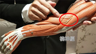
Located 10 centimeters above the lateral epicondyle of the humerus, the radial nerve is often wrapped by intersecting tendinous muscles, like a tunnel through which a train passes. Here, the radial nerve is also prone to compression, especially after intense upper limb activity (such as playing tennis, baseball, etc.), which can easily damage the nerve.
Manual release or acupuncture and moxibustion can be performed about 10 cm above the lateral epicondyle of the humerus.
PS: Of course, the area near the lateral epicondyle of the humerus is also an important node, just like a train platform.
9. Dorsal myotube.
The deep branch of the radial nerve (posterior interosseous nerve) passes through a muscular gap in the retrospinatus muscle, which forms an arc-shaped distal tendon arch (Frohse arch) at the starting point of the lateral and medial epicondyle of the humerus. This arch can cause compression of the posterior interosseous nerve at this location.
Continuing downwards, the superficial branch (cutaneous branch) of the radial nerve turns to the dorsal side at the junction of the middle and lower one-third of the forearm, and continues downwards to the back of the hand, distributed on the radial half skin of the back of the hand and the dorsal skin of the three and a half fingers near the radial side.
If the radial nerve is compressed and damaged, sensory disorders will occur in the skin area it controls, especially in the "tiger mouth area" skin.
Of course, after injury, weakness in the dominant muscles is also a typical symptom, such as the brachioradialis, triceps, retrospinatus, and other forearm extensor muscles. Muscle strength testing is also a good way to check for nerve damage.
A typical indication is paralysis of the extensor muscles of the forearm, manifested as a "wrist hanging" state when lifting the forearm.
10. Intersection of the middle and lower one-third of the forearm.
As mentioned earlier, the superficial branch (cutaneous branch) of the radial nerve turns to the dorsal side at the junction of the middle and lower one-third of the forearm. At this bend, it is like a winding mountain road, and it is also easy to get stuck.
We have found in clinical practice that this place is often related to a traditional Chinese medicine acupoint, and stimulating this acupoint can indeed be helpful. This acupoint is called the Lieque acupoint.
How about it, the anatomy of modern medicine and the acupuncture points of traditional Chinese medicine sometimes have intricate and magical connections, which we propose for everyone's reference.
Can you remember all ten points clearly?