What is patellar softening?
To know what disease this is, you need to first understand what patella is.
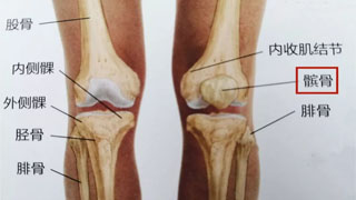
There is a circular bone in front of our knee. We can all touch it, feel it. If you stretch your leg straight and relax, you can move this bone left and right, and this bone is the patella. We know that the famous military strategist Sun Bin in the early Warring States period (who pioneered classic tactics such as besieging Wei to save Zhao and besieging cities for reinforcements) had his patella removed from both legs by his opponent Pang Juan, resulting in lifelong disability and renaming him Sun Bin.
The patella is a bone embedded in the quadriceps tendon, serving as a fulcrum for the quadriceps muscle to exert force. It is crucial for the normal function of the knee joint, and about a quarter of knee joint pain is caused by patellar lesions.
This is a side view of our knee joint skeleton. The independent bone in front is the patella, and the yellow circle below the patella is the cartilage of the patella. It can be seen that these cartilages are the contact surfaces between different bones, playing a role in reducing friction. And the image on the right shows that the cartilage has degenerated and softened, causing pain.
Patellar chondromalacia
Patellar chondromalacia is a condition characterized by softening and degeneration of the articular cartilage of the patella. Patellofemoral chondromalacia is sometimes considered synonymous with patellofemoral pain syndrome, but the term patellofemoral pain syndrome only applies to individuals without cartilage damage.
Patellar chondromalacia is considered the most common cause of chronic knee pain.
The cartilage beneath the patella or knee bone acts as a natural shock absorber. Overuse, injury, and many other factors can lead to cartilage deterioration and damage.
Cartilage softening is common among young athletes, but it can also occur in elderly people with knee arthritis. There is a preference for women.
The causes of patellar chondromalacia
Patellofemoral chondromalacia occurs due to a disruption in the biomechanical relationship of the patellofemoral joint. The patella tilts outward or subluxates, causing the cartilage on the inner side of the patella to collide with the trochanter of the femoral condyle, resulting in excessive wear of the cartilage in the lateral joint space, loss of chondrocytes, bone hyperplasia, and a series of pathological changes such as narrowing of the joint space. Various clinical symptoms appear: pain in the anterior side of the knee joint, aggravated pain when standing up or going downstairs or downhill for prolonged sitting, often accompanied by leg weakness, joint coldness, or repeated swelling and fluid accumulation in the knee joint. In severe cases, osteoarthritis can develop, so early diagnosis and appropriate prevention and treatment measures are crucial to prevent its occurrence or development.
Usually, the quadriceps pull the patella in a straight line through the end of the femur. Patients with patellar chondromalacia often experience abnormal trajectory of the patella towards the lateral femur, leading to femoral friction and chronic inflammation and pain. Abnormal patellar movement caused by improper functioning of bones, tendons, or ligaments.
Actually, it is called patellar chondromalacia, and its full name is patellar chondromalacia. We have all eaten pig bones, and pig bones have "brittle bones", which are cartilage. They are very smooth and serve as the friction surface of our joints. Long term friction can lead to cartilage degeneration or damage, and this degeneration is also graded. The earliest is cartilage softening, also known as cartilage softening. If degeneration continues, the cartilage becomes thinner, and then it falls off. So, cartilage softening is an early stage of cartilage degeneration, and there is still hope!
Risk factors for patellar chondromalacia
Age
Teenagers and young adults are at high risk as rapid muscle and bone development during growth spurt can lead to short-term muscle imbalance.
Flat feet
Flat feet can bring greater pressure to the knee joint.
trauma
Previous injuries, such as patellar dislocation or subluxation, increase the risk of patellar chondromalacia.
Chronic friction between the patella and femoral groove can also lead to this condition.
activity
High levels of activity or pressure on the knee joint can increase the risk of knee problems.
Knee joint pathology
Inflammation such as arthritis can prevent the normal functioning of the knee bones. Other knee joint diseases that can cause patellar chondromalacia include high patella, quadriceps imbalance, hamstring weakness, congenital abnormalities, poor patellar trajectory, synovial folds, tight iliotibial tract, neuroma, bursitis, etc.
overuse
People engaged in football, gymnastics, cycling, rowing, tennis, ballet, basketball, volleyball, running, combat sports, snowboarding, skateboarding, and even swimming face higher risks.
Clinical manifestations of patellar chondromalacia
People with anterior knee pain, especially adolescent women or young adults, should be suspected of having patellar chondromalacia.
Patients with patellar chondromalacia usually present with pain in the anterior part of the knee joint or mild discomfort on the medial side of the knee joint, which can worsen with activities such as running, jumping, climbing mountains, or going down stairs, or prolonged sitting. Knee pain may occur after kneeling, squatting, or sitting for a long time.
Patients may also complain of stiff or tight knees. Examination may reveal mild swelling in the knee.
Imaging Science
Knee radiographs cannot evaluate cartilage changes and can only display bone changes (if present), such as osteoarthritis.
MRI is the preferred method for evaluating patellar cartilage and can display cartilage abnormalities.
Grading of patellar chondromalacia
class a
Softening of cartilage in the knee area.
Level 2
Cartilage softening and abnormal surface features. This usually indicates the beginning of organizational erosion.
Level 3
Cartilage thinning and tissue activity deterioration.
Level 4
Significant loss of cartilage accompanied by bone exposure
Differential diagnosis
Patellar tendon degeneration
Sindin Larson Johnson disease
Patellar sleeve fracture
Treatment of patellar chondromalacia
Rice therapy
Reduce activities
Non steroidal anti-inflammatory drugs
practice
Fixed bicycle
Swimming
Stretching and strengthening the quadriceps, especially the medial thigh muscles
Cardiovascular regulation
The correct training plan
Suitable shoes
Through appropriate treatment, patients can usually recover their full functional levels.
When the imbalance is severe or the patient does not show improvement, surgical selection may be necessary
Arthroscopic debridement and irrigation
Joint surface repair
Instability correction
Patellar resection surgery
prevention
Avoid repeated pressure on the knee bones.
Use knee pads
Strengthening exercises for quadriceps and hamstring muscles, as well as abductors and adductors.
Wear insoles for correcting flat feet
Maintain an appropriate weight