Learning anatomy is crucial for rehabilitation therapy personnel. Taking the radial nerve as an example, this article analyzes its related knowledge.
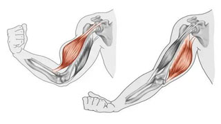
The radial nerve is prone to fracture, excessive callus, radial head dislocation, or surgical damage in the middle and lower third of the humerus. It originates from the nerve root fibers of the fifth to eighth cervical segments and the first thoracic segment, and is a continuation of the posterior bundle of the brachial plexus nerve. It exits from the upper arm axilla, travels along the deep brachial artery, passes through the gap between the long and medial heads of the triceps brachii to the dorsal side of the upper arm, and then descends along the radial nerve groove of the humerus between the inner and outer heads of the triceps brachii. It branches into shallow and deep branches above the elbow joint and enters the forearm, innervating muscles such as the triceps brachii along the way.
Learning neurological knowledge is because all structures are innervated by nerves, and problems in the body may be accompanied by neurological problems, and treatment can also affect the nerves. The radial nerve is the thickest, longest, and largest branch of the brachial plexus. The brachial plexus is mainly composed of the anterior branches of the 5th to 8th cervical nerves and the anterior branch of the first thoracic nerve, with numerous branches that can be memorized as "five/three/six/three/fourteen branches".
The compression points of the radial nerve include:
1. Cervical spine: Misalignment of the cervical spine can easily cause compression of the cervical or brachial plexus nerves, and cervical spine reduction and training should be considered during rehabilitation.
2. Diagonal muscle: Long term lowering of the head causes tension in the diagonal muscle, which compresses the brachial plexus nerve. The release technique is performed on the lateral side of the cervical spine.
3. Pectoral muscles: Long term sitting at the desk causes anterior rotation of the shoulders, and the pectoral muscles tense and compress nerves. The focus of the technique is on the coracoid process and release to the rib surface.
4. Subclavicular muscle: may compress the radial nerve.
5. At the corner of the armpit and arm: posture issues can easily cause jamming and compression.
6. Lower three sided hole: easy to get stuck and compressed near the nerve outlet.
7. Radial nerve groove: Lying sideways can easily damage the upper limbs, also known as "honeymoon syndrome".
8. Penetrating the lateral muscular septum: the upper limb is easy to be compressed after high-intensity exercise, which can be released or acupuncture and moxibustion at 10 cm above the lateral epicondyle of the humerus. The vicinity of the lateral epicondyle of the humerus is also an important node.
9. Dorsal myotube: A tendinous arch formed at the origin of the supine muscle is prone to compression of the posterior interosseous nerve.
10. Intersection of the middle and lower one-third of the forearm: This area is prone to getting stuck and compressed at the bend, which is related to the missing acupoints in traditional Chinese medicine.
Damage to the radial nerve can cause sensory disorders in the innervated skin, particularly in the "tiger mouth area", and can also lead to muscle weakness in the innervated muscles, such as paralysis of the forearm extensor muscles in a "wrist hanging" state. Muscle strength testing can detect nerve damage.